
Bladder tumours: endoscopic treatment and robot-assisted surgery
Blood in the urine is most often the first sign of a bladder tumour. Depending on how deep it goes, treatment ranges from endoscopic resection (TURBT) to robot-assisted cystectomy. Diagnosis, treatments and follow-up, explained by Dr Najib Abakka in Rabat.
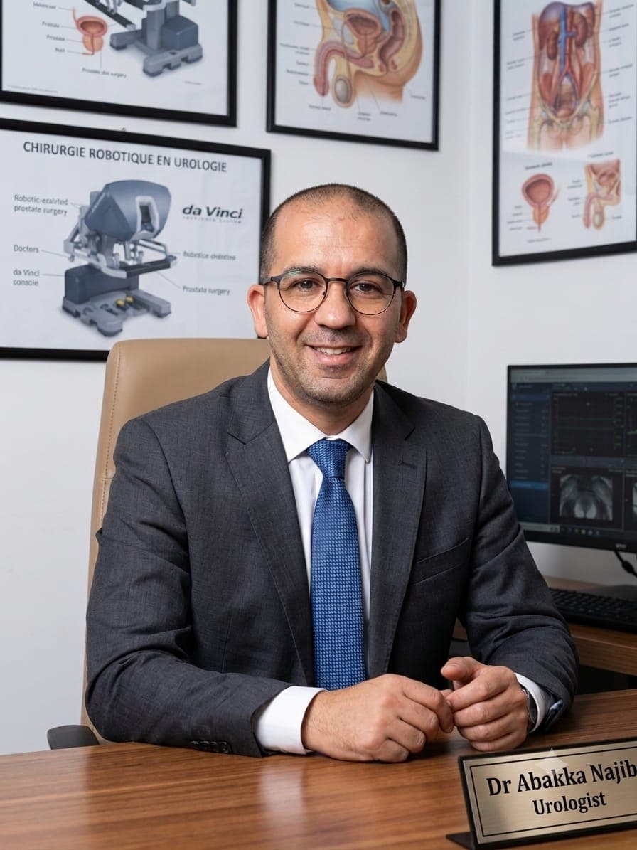
Dr Najib Abakka
Urological Surgeon in Rabat
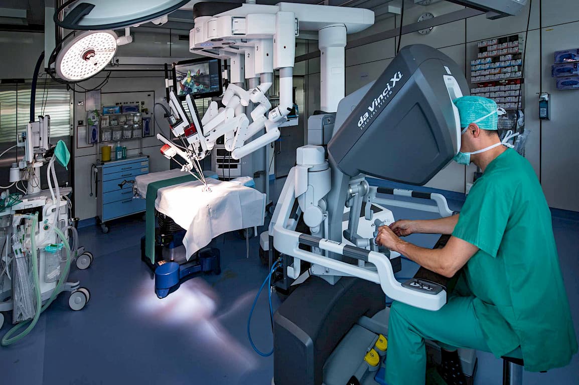
La cystectomie radicale réalisée avec l'assistance du robot, par voie mini-invasive.
Understanding bladder tumours
The bladder is the reservoir that stores urine produced by the kidneys before it is evacuated through the urethra. Its wall is lined with an inner coating, the urothelium, and includes deeper down a muscle layer that contracts to empty the bladder.
A bladder tumour most often arises from this inner lining. Two very different situations are distinguished, and they determine the entire treatment:
The superficial tumour (not invading the muscle): it remains limited to the inner lining and the layer just below it. It is the most frequent form at the time of diagnosis.
The invasive tumour: it has crossed the wall and reached the bladder muscle, sometimes beyond. It requires radical treatment.
Signs that should alert you
Blood in the urine (haematuria), visible to the naked eye, often painless and intermittent: this is the most frequent sign.
Frequent or urgent urges to urinate, burning on urination, which can wrongly suggest a recurring urinary infection.
Later: pelvic pain, lower back pain or unexplained fatigue.
Do not wait
Haematuria, even a single episode and even if it disappears on its own, always justifies a urological work-up. Caught early, a bladder tumour is most often treated while preserving the bladder.
Risk factors — Smoking is the main risk factor: the breakdown products of smoke are eliminated in the urine and remain in prolonged contact with the bladder wall. Next come occupational exposure to certain chemicals (dyes, rubber, paint, leather and aluminium industries) and, more rarely, chronic irritation of the bladder. Stopping smoking reduces the risk of recurrence and is an integral part of management.
Diagnosis
The work-up combines clinical examination, imaging tests and above all direct exploration of the bladder:
Cystoscopy (bladder endoscopy): a thin flexible optical tube is introduced through the urethra, under local anaesthesia, to visualise the inside of the bladder directly and locate the tumour.
Imaging: ultrasound of the urinary tract, then CT (CT urography) to assess the whole urinary tract and look for spread.
Urine cytology: looking for abnormal cells in the urine.
Histopathological analysis: microscopic examination of the tissue removed during resection confirms the diagnosis and clarifies whether the muscle is involved — it is this analysis that decides what follows.
Endoscopic treatment: transurethral resection (TURBT)
When the tumour is superficial, it is removed through the natural passages, without any incision: this is transurethral resection of the bladder (TURBT). It is both the procedure that confirms the diagnosis and the first treatment.
The procedure takes place under general or locoregional anaesthesia (spinal anaesthesia), in the operating theatre.
The surgeon introduces a rigid endoscope (resectoscope) through the urethra. Under direct vision, the tumour is cut into small fragments using an electric loop, including a sample of the muscle underneath: this sample is what allows verification that the tumour does not infiltrate the wall.
The removed fragments are sent to the histopathology laboratory. The result, available within a few days, clarifies the nature of the tumour, its grade and its depth.
A urinary catheter is left in place for one to a few days, until the urine becomes clear again. The hospital stay is short and return to activities is fast.
After resection: intravesical instillations — Superficial tumours tend to recur. To reduce this risk, a treatment can be instilled directly into the bladder through a catheter, during consultations, in repeated sessions:
Intravesical chemotherapy (for example mitomycin), often started very soon after resection for low- or intermediate-risk tumours.
Immunotherapy with BCG, for high-risk superficial tumours: it stimulates the bladder's local defences and reduces both recurrence and the risk of progression to an invasive form.
Regular cystoscopy follow-up is then essential, because a recurrence detected early can again be treated endoscopically.
Radical treatment: robot-assisted cystectomy
When the tumour invades the bladder muscle — or in certain very high-risk superficial forms that escape conservative treatments — the reference treatment is complete removal of the bladder: radical cystectomy. In our practice, it is performed with robotic assistance, by a minimally invasive approach.
Superficial tumour
Resection through the urethra (TURBT), without incision
The bladder is preserved
Instillations (mitomycin or BCG) according to risk
Regular cystoscopic follow-up
Invasive tumour
Radical cystectomy: removal of the bladder
Associated pelvic lymph node dissection
Urinary diversion reconstructed at the same time
Complete analysis of the surgical specimen
How robot-assisted cystectomy proceeds — Under general anaesthesia, small incisions of a few millimetres allow the 3D camera and the robot's instruments to be introduced. There is no large opening of the abdomen.
Seated at the console, the surgeon removes the bladder. In men, the prostate and seminal vesicles are removed at the same time; in women, removal generally includes the uterus and part of the vagina, depending on the extent of the tumour.
The lymph nodes of the pelvis are removed: they are analysed to clarify the exact stage of the disease and detect any microscopic spread.
The urine must then be diverted: the surgeon performs, during the same operation and also with the robot, the diversion chosen with the patient beforehand (see below).
All removed tissue is examined in the laboratory. This result defines the definitive stage and the possible need for additional treatment.
What the robot brings to the patient
Magnified 3D vision, highly precise articulated instruments, movements filtered of tremor: compared with open surgery, the robot-assisted approach reduces bleeding, postoperative pain and scarring, and facilitates faster recovery — with the same demands of oncological radicality: removing the whole tumour with healthy margins and a complete lymph node dissection.
In some cases, chemotherapy may be offered before the operation (called neoadjuvant) or afterwards, in multidisciplinary discussion with the oncologist, depending on the tumour stage and the patient's general condition.
After bladder removal: how is urine diverted?
With the bladder removed, a new route must be created to evacuate the urine produced by the kidneys. The choice of diversion is discussed with the patient before the operation, depending on the tumour, the patient's condition and wishes.
The most frequent — Ileal conduit (Bricker procedure) — A short segment of small intestine is used as a conduit: the ureters are connected to it, and its end opens onto the skin of the abdomen (stoma). Urine flows continuously into a collection bag worn discreetly under clothing. It is the simplest and most time-tested diversion.
The bag-free alternative — Orthotopic neobladder — A segment of intestine is remodelled into a reservoir shape and connected to the urethra, in place of the former bladder. The patient then urinates through the natural passages, without an external bag. It requires training (regular emptying, sometimes self-catheterisation) and cannot be offered in all situations.
In particular cases — Cutaneous ureterostomy — The ureters are connected directly to the skin, with a collection bag worn. A simpler procedure, reserved for frail patients or particular situations.
Whatever the diversion, support is organised: stoma care training if needed, practical advice and close follow-up in the first few weeks.
Postoperative recovery and follow-up
After endoscopic resection (TURBT) — The urinary catheter is removed after one to a few days. Pink urine and transient urgent urges are frequent and fade within one to two weeks. Return to work is fast. Follow-up then rests on regular cystoscopies, the frequency of which depends on the risk of recurrence.
After robot-assisted cystectomy — Hospital stay generally lasts several days. Eating and walking are resumed early, which promotes recovery. Drains and catheters are removed gradually, and mastery of the urinary diversion is learned before discharge. Recovery continues at home, with a gradual return to activities over several weeks.
Long-term follow-up — After treatment of a bladder tumour, long-term follow-up is essential: regular consultations, imaging and tests to detect recurrence or spread, monitor kidney function and the state of the diversion. Stopping smoking remains, at every stage, one of the most effective measures to protect what follows.
Frequently asked questions
Does blood in the urine necessarily mean cancer?
No: a urinary infection, a stone or an enlarged prostate can also be the cause. But only a urological consultation with cystoscopy can settle the matter. All haematuria must therefore be investigated, even if it happens only once.
Is endoscopic resection painful?
The procedure itself takes place under general or locoregional anaesthesia: the patient feels nothing. In the days that follow, burning on urination and frequent urges are possible, then gradually ease.
Can a bladder tumour be cured while keeping the bladder?
Yes, when the tumour is superficial: endoscopic resection, combined if needed with BCG or chemotherapy instillations, allows it to be controlled while preserving the bladder, at the cost of regular follow-up. Removal of the bladder is only proposed when the tumour invades the muscle or presents a high risk of doing so.
Does the robot operate on its own?
No. The robot is a tool entirely controlled by the surgeon, seated at a console a few metres from the patient. Every movement is performed by the surgeon: the robot transmits the surgeon's movements to the instruments, refining them, with magnified 3D vision of the operated area.
How does one urinate after bladder removal?
Urine is diverted either to a collection bag worn on the abdomen (Bricker ileal conduit, the most frequent), or to a neobladder reconstructed from a segment of intestine and connected to the urethra, which allows urination through the natural passages. The choice is discussed before the operation.
Is chemotherapy always necessary?
No. It depends on the tumour stage and the results of tissue analysis. It may be offered before cystectomy (neoadjuvant), after the operation, or as local instillations into the bladder for superficial tumours. The decision is taken at a multidisciplinary team meeting.
Related guides & articles
Browse all articles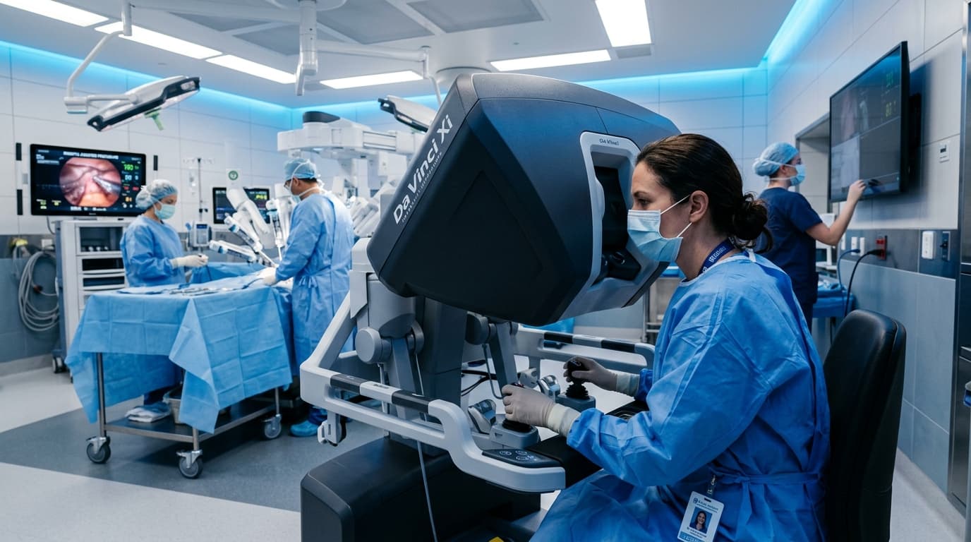
Robotic Surgery in Urology in Rabat: What Patients Should Know Before Deciding
Understand robot-assisted urological surgery, its clinical indications, limits, and essential questions to ask before deciding.
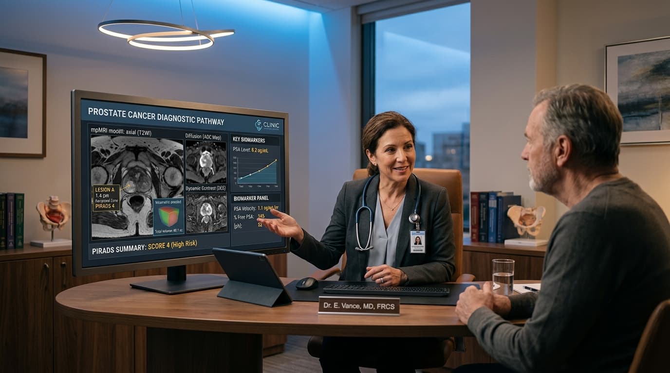
Prostate Cancer: From PSA to MRI, Understanding the Diagnostic Pathway Before Discussing Surgery
An elevated PSA does not automatically mean cancer. Learn how PSA, clinical exam, multiparametric MRI, and targeted biopsy fit together.
Robot-Assisted Radical Prostatectomy: 12 Essential Questions to Ask Before Surgery
Facing prostatectomy? Here are 12 vital questions concerning surgical indication, nerve sparing, continence, erectile function, and follow-up.