
Female Genital Prolapse: Treatment by Sacrocolpopexy (Promontofixation)
Pelvic organ prolapse in women: understanding genital prolapse (cystocele, rectocele, uterine prolapse), its symptoms, its diagnosis, and laparoscopic sacrocolpopexy, the reference surgical treatment for the apical compartment.

Dr Najib Abakka
Urological Surgeon in Rabat
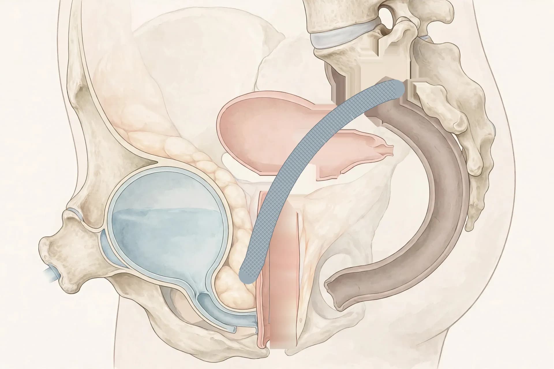
Schéma anatomique du plancher pelvien féminin : le vagin est soutenu et fixé au promontoire sacré par une bandelette.
What is genital prolapse?
Genital prolapse — sometimes called "pelvic organ prolapse" — occurs when one or more pelvic organs descend and press on the wall of the vagina, or even protrude through the vaginal opening. The organs most often involved are the bladder, the uterus and the rectum.
The pelvic floor is the set of muscles and ligaments that support these organs, like a hammock. When this support weakens, the organs slip downwards. It is a common problem, particularly after the menopause, and there is no reason to feel ashamed of it or to consider it inevitable: solutions exist, from rehabilitation to surgery.
The different types of prolapse
Prolapse is described according to the organ that descends:
Cystocele: the bladder descends and presses on the front wall of the vagina.
Rectocele: the rectum descends and presses on the back wall of the vagina.
Uterine prolapse: the uterus itself descends into the vagina.
Vaginal vault prolapse: after a hysterectomy (removal of the uterus), the top of the vagina, known as the "vault", descends.
Enterocele: rarer, this is the small intestine descending between the vagina and the rectum.
It is common for several compartments to be affected at the same time: this is called multi-compartment prolapse.
What are the symptoms?
Symptoms vary from one woman to another, and they are not always proportional to the extent of the descent. The most common are:
A heaviness in the lower abdomen or a dragging sensation downwards.
A sensation of a bulge in the vagina, or the feeling that "something is coming out", especially when standing, when walking or at the end of the day. This discomfort generally improves when lying down.
Urinary symptoms: urgent need to urinate, leakage on effort (coughing, laughing, lifting), difficulty urinating, a feeling of incomplete bladder emptying, recurrent urinary infections.
Bowel symptoms: constipation, a feeling of incomplete rectal emptying, needing to press with the fingers to complete a bowel movement.
Discomfort or pain during sexual intercourse, or apprehension about it.
Sometimes lower back or pelvic pain, especially at the end of the day.
Why does it happen? Causes and risk factors
Prolapse most often results from a gradual weakening of the pelvic floor muscles and ligaments. The main risk factors are:
Vaginal deliveries, especially when they are numerous, when the baby is of high birth weight, or when the delivery was difficult.
Age and the menopause: falling oestrogen levels make the supporting tissues more fragile.
Overweight and obesity, which increase pressure inside the abdomen.
Chronic constipation and repeated straining.
Chronic cough.
Repeated heavy lifting.
A family predisposition: some women naturally have less resistant tissues.
A history of pelvic surgery, notably hysterectomy.
How is it diagnosed?
Diagnosis is made during a consultation, in two steps. First, the doctor listens to the symptoms and their impact on daily life. Then, a clinical examination is performed: a gynaecological examination, at rest and then asking the patient to bear down (Valsalva manoeuvre), to see which organs descend and how far.
To describe the extent of the descent precisely and reproducibly, an international classification is used, the POP-Q classification. Simply put, it has five stages: stage 0 means there is no prolapse; stage I corresponds to a slight descent; stage II, to a descent reaching the level of the vaginal opening; stage III, to a descent going beyond the vaginal opening; stage IV, to complete protrusion.
Depending on the symptoms, additional investigations may be offered: urodynamic testing (which measures how the bladder works) in case of urinary symptoms, an ultrasound scan, and screening for so-called "occult" urinary incontinence — that is, incontinence hidden by the prolapse itself, which may become apparent once the prolapse is corrected. Before any surgery, preoperative blood tests and an anaesthetic consultation are also carried out.
Treatments without surgery
When symptoms are moderate, or while a surgical decision is pending, several conservative treatments can help:
Pelvic floor rehabilitation: exercises guided by a physiotherapist to strengthen the pelvic floor muscles, learn how to bear down correctly, and protect the perineum in everyday movements.
A pessary: a small silicone device placed in the vagina, which supports the organs and holds them in place. It is removable, must be fitted to each patient, and requires regular follow-up.
Lifestyle measures: weight loss in case of overweight, treatment of constipation and cough, avoiding heavy lifting.
Local oestrogen treatment, after the menopause, may be offered by the doctor to improve the quality of the vaginal tissues.
When is surgery considered?
Surgery is considered when symptoms interfere with daily life despite conservative treatments, when a pessary is poorly tolerated or insufficient, or when the prolapse is severe. It is always the patient who decides, after receiving full information about the expected benefits and the risks of each option. There is no urgency to operate on a prolapse, except in rare cases: there is time to think it over.
Sacrocolpopexy: what does it involve?
Sacrocolpopexy is an operation that "reattaches" the top of the vagina — or the cervix when the uterus is preserved — to a solid bony landmark in the pelvis, the sacral promontory, that is, the front part of the sacrum, deep in the pelvis. Fixation is achieved using a strip of synthetic material (polypropylene mesh), which in a way takes over the role of the weakened ligaments and restores the vagina to its natural position and axis.
It is called sacrocolpopexy when the vagina is fixed after a hysterectomy, and sacrohysteropexy when the uterus is preserved and fixation is made to the cervix.
Why is sacrocolpopexy the reference treatment for the apical compartment?
The apical compartment, the top of the vagina, is in a way the "keystone" of the pelvic floor. When it descends, it often drags the other compartments down with it. That is why solidly correcting this compartment is essential, and why sacrocolpopexy is the reference treatment for it.
Its advantages are as follows: durable anatomical results, because fixation to the promontory is solid; restoration of the natural depth and axis of the vagina; the possibility of treating the anterior (bladder) and posterior (rectum) compartments at the same time; and preservation of sexual function, because unlike some vaginal approaches, it does not narrow the vagina. Finally, it is performed through small incisions, by laparoscopy.
How does the operation unfold?
The operation is performed under general anaesthesia, by laparoscopy: the surgeon works through a few small incisions in the abdomen, guided by a camera. He first puts the organs back in place, then fixes the top of the vagina — or the cervix when the uterus is preserved — to a solid landmark of the pelvis, the sacral promontory, using a thin synthetic mesh strip. This strip takes over the role of the weakened ligaments and holds the organs in their natural position, without narrowing the vagina.
The same operation can also be performed with robotic assistance, on the same principle.
The operation generally lasts 2 to 3 hours, and the hospital stay is generally 1 to 2 days.
What are the possible risks and complications?
Like any surgery, sacrocolpopexy carries risks, which the surgeon explains in consultation before the operation:
The risks common to any surgery: bleeding, infection, and, rarely, injury to a neighbouring organ (bladder, rectum, ureters, blood vessels).
Mesh exposure in the vagina (sometimes called erosion): the mesh may become visible or palpable in the vagina, which may require local treatment or, more rarely, further surgery.
Pain during sexual intercourse (dyspareunia) or persistent pelvic pain.
Bowel symptoms, notably constipation.
Urinary incontinence appearing or persisting: incontinence previously hidden by the prolapse may become apparent once the prolapse is corrected.
Recurrence of the prolapse or descent of another compartment over time.
Conversion to open surgery: rarely, if technical difficulties arise, the surgeon may decide to switch to a conventional incision to complete the operation safely.
After the operation: recovery and convalescence
Recovery is generally straightforward. The urinary catheter is generally removed the day after the operation. Walking is resumed the same day or the next day. Pain is generally moderate and relieved by painkillers. Light vaginal bleeding may persist for a few days.
Activities are resumed gradually. Heavy efforts and heavy lifting should generally be avoided for 6 weeks, and sexual intercourse should generally be avoided for about 6 weeks, as advised by the surgeon. Follow-up consultations allow healing of the vagina and the correct position of the organs to be checked.
When should you seek medical advice?
You should seek medical advice if you feel pelvic heaviness, a sensation of a bulge in the vagina, urinary or bowel symptoms, or discomfort during intercourse. Do not wait for the prolapse to get worse: a consultation allows the situation to be assessed and an appropriate solution to be offered, whether conservative or surgical.
A treatment chosen for each patient
The choice of treatment — rehabilitation, pessary or surgery, and the type of operation — is always individualised: it is decided after a full examination, taking into account your symptoms, your age, your state of health, your medical history and your expectations. If you recognise yourself in the symptoms described in this article, a consultation allows you to take stock and discuss the solution best suited to your case.
---
Related guides & articles
Browse all articles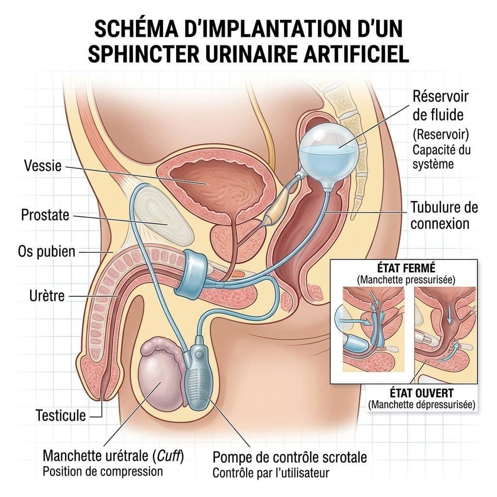
Artificial urinary sphincter: treating severe urinary incontinence in men
After prostate surgery, particularly prostatectomy, some men have persistent, severe stress urinary incontinence. When pelvic floor rehabilitation and other treatments are not enough, the artificial urinary sphincter is the reference treatment for severe incontinence. This fully implanted device keeps the urethra closed at rest and opens on demand, by simply pressing a pump, to allow urination. The actual indication is decided in consultation, after quantifying the incontinence and assessing bladder function.
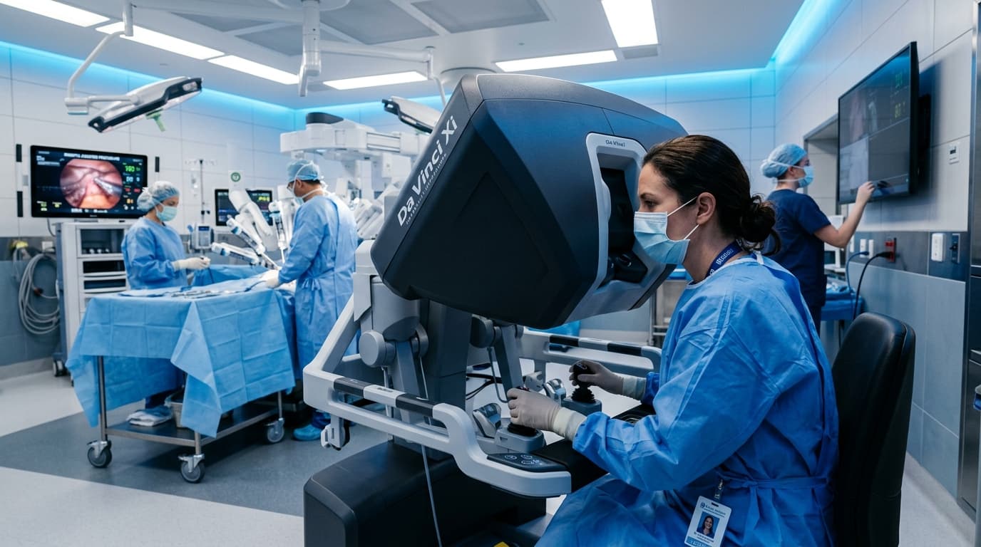
Robotic Surgery in Urology in Rabat: What Patients Should Know Before Deciding
Understand robot-assisted urological surgery, its clinical indications, limits, and essential questions to ask before deciding.

Prostate Cancer: From PSA to MRI, Understanding the Diagnostic Pathway Before Discussing Surgery
An elevated PSA does not automatically mean cancer. Learn how PSA, clinical exam, multiparametric MRI, and targeted biopsy fit together.