
Kidney tumours: partial or total nephrectomy with robotic assistance
A localised kidney tumour is most often treated with surgery: partial nephrectomy to preserve the kidney, or total nephrectomy when that is not possible, with robotic assistance. How the choice is made, what the operation involves and what follows, explained by Dr Najib Abakka in Rabat.

Dr Najib Abakka
Urological Surgeon in Rabat
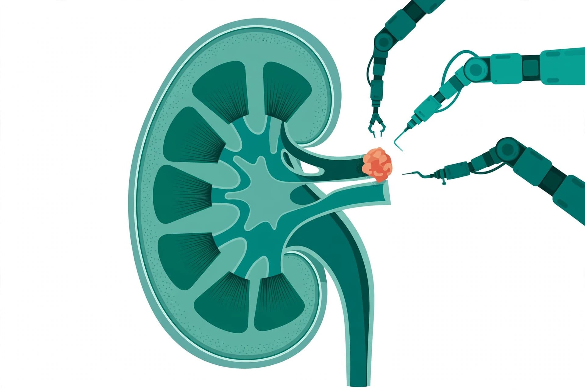
Le robot permet souvent de n'enlever que la tumeur et de conserver le reste du rein.
What is a kidney tumour?
A kidney tumour is a mass that develops in kidney tissue. Around 90% of kidney cancers are renal cell carcinomas, the most frequent form of which is clear cell carcinoma. Not all kidney masses are cancers: some are benign (cyst, angiomyolipoma, oncocytoma), but a solid mass that enhances with contrast on CT must be considered suspicious until proven otherwise.
Most often found by chance — Most kidney tumours are discovered by chance, during an ultrasound or CT scan performed for another reason (abdominal pain, a check-up, follow-up). This is relatively good news: these incidental tumours are often small and localised, and therefore more amenable to kidney-sparing surgery.
Symptoms, when there are any
Blood in the urine (haematuria), visible or detected on testing
Flank or back pain, on one side only
A palpable mass in the flank
Sometimes: fatigue, weight loss, persistent fever, anaemia
The classic triad (blood, pain, mass) is rare nowadays: it often corresponds to tumours that are already advanced. The absence of symptoms does not rule out a tumour.
Risk factors
Smoking — the most important avoidable factor
Obesity
High blood pressure
Chronic kidney disease and long-term dialysis
Occupational exposures (certain solvents, cadmium)
Rare hereditary forms (von Hippel-Lindau disease, etc.)
The principle that guides everything else: remove the tumour completely, while preserving as much healthy kidney as possible — without ever compromising the anti-cancer result.
Imaging, kidney function and staging
Before any decision, three questions must be answered: is the tumour localised? What is its exact size and position within the kidney? And how are both kidneys working? Each case is then discussed at a multidisciplinary team meeting (MDT) — urologist, radiologist, oncologist, histopathologist — before a treatment is proposed to you.
Imaging: CT and MRI
Contrast-enhanced CT of the abdomen and pelvis (CT urography): the central examination. It measures the tumour, clarifies its relationship with blood vessels and the urinary tract, and looks for spread.
Chest CT: for staging in at-risk forms.
Kidney MRI: as a complement or an alternative — allergy to contrast medium, doubt about the nature of the mass, involvement of the renal vein or vena cava, impaired kidney function.
Imaging also allows calculation of the complexity scores (see below) that help predict the difficulty of partial surgery.
Kidney function
Creatinine and glomerular filtration rate (eGFR): to know how your kidneys are working overall before the operation.
Renal scintigraphy (if needed): it measures each kidney's share of the work separately — useful when deciding between partial and total surgery, or when one kidney is already working poorly.
General work-up: blood count, anaesthesia assessment, heart and lung function depending on your situation.
TNM staging: size matters — For a tumour confined to the kidney (no lymph nodes or metastases), the stage depends mainly on size. Two thresholds structure the decision: 4 cm and 7 cm.
What about kidney biopsy? It is not routine — Unlike prostate cancer, not all kidney masses are biopsied. Percutaneous kidney biopsy (under CT or ultrasound guidance) is reserved for selected indications: when its result would change the strategy — doubt about a benign lesion or another disease (lymphoma, metastasis from another cancer, infection), a small mass in a frail patient for whom surveillance or ablative treatment is being considered, or metastatic tumour before medical treatment. When surgery has already been decided and will be needed in any case, analysis of the surgical specimen gives the definitive diagnosis.
Partial or total: how is the decision made?
Both operations have the same goal — curing the cancer — and, at the same stage and when a partial nephrectomy can be performed under good conditions, the anti-cancer results are comparable. The difference lies in the kidney function preserved and in the technical complexity. Click on each procedure to see it in detail.
The tumour is removed, the kidney is kept — The surgeon removes only the tumour, surrounded by a thin margin of healthy tissue, then reconstructs the remaining kidney. It is the reference treatment for T1a tumours (≤ 4 cm) and is preferred for T1b tumours whenever complexity allows.
Its advantages
Preserves kidney function: lower risk of chronic kidney disease, long-term dialysis and associated cardiovascular events
Anti-cancer results equivalent to total nephrectomy for well-selected localised tumours
A valuable kidney "reserve" in case of a tumour on the other side, one day
Its requirements
More complex operation: the artery must be clamped, the tumour removed, then the kidney reconstructed (renorrhaphy) within a limited time
Specific risk of urine leak and secondary bleeding, low but real
Not always possible: central tumour, very large tumour, or one invading blood vessels
The whole kidney is removed, with the tumour — The surgeon removes the entire affected kidney, surrounded by its fat, after ligating the artery, the vein and the ureter. This is the operation indicated when safe partial surgery is not feasible.
Its main indications
Large tumour (often > 7 cm) or occupying most of the kidney
Central / hilar tumour, in contact with the large blood vessels
Venous invasion (thrombus in the renal vein or vena cava)
Multifocal tumour, or a kidney that is already non-functioning
What you should know
You can live perfectly normally with a single kidney, if the other one works well
Technically simpler operation, often faster than a partial nephrectomy
Overall kidney function decreases: long-term monitoring, protection of the remaining kidney (blood pressure, diabetes, medicines)
By comparison, partial nephrectomy removes only the tumour with a margin of healthy tissue, then reconstructs the remaining kidney; it is indicated for tumours of 4 cm or less (T1a), for T1b tumours (4 to 7 cm) when feasible, and in all imperative situations. Total nephrectomy removes the whole kidney with its surrounding fat, after dividing the artery, vein and ureter; it is indicated for tumours larger than 7 cm, central or hilar tumours, venous invasion, and destroyed or non-functioning kidneys.
After a partial nephrectomy, kidney function is preserved as much as possible because the healthy tissue keeps filtering; after a total nephrectomy, it relies on the remaining kidney alone, with a drop in eGFR to monitor. Partial nephrectomy is technically more demanding (clamping, excision and renorrhaphy under ischaemia time pressure), while total nephrectomy is more standardised, with an added vascular procedure if there is a venous thrombus. The specific risks of partial nephrectomy are secondary bleeding, urine leak (fistula) and pseudoaneurysm; total nephrectomy carries fewer specific local risks, but a greater overall renal impact.
Against cancer, results are equivalent at the same stage when the tumour is removed completely with healthy margins: what matters is the quality of the excision, not the amount of kidney removed.
"Imperative" partial nephrectomy: there is no choice
Single kidney (anatomical or functional)
Bilateral tumours
Pre-existing chronic kidney disease
Hereditary disease with a risk of repeated tumours
"Elective" partial nephrectomy: it is chosen
RENAL and PADUA scores: measuring complexity before operating
How the robotic operation proceeds
The robot decides nothing: it is entirely controlled by the surgeon, seated at a console. It transmits the surgeon's movements while removing tremor, with articulated instruments that rotate like a human wrist, and a 3D camera magnified up to 10 times. For the kidney — a deep, highly vascular organ that often has to be reconstructed — these advantages matter particularly.
Transperitoneal approach — The most widely used. The instruments pass through the abdominal cavity, which gives a large working space and familiar landmarks. It is preferred for anterior tumours, large tumours, or when a lymph node dissection or an associated procedure is planned.
Retroperitoneal approach — The instruments pass behind the abdomen, directly onto the kidney, without crossing the peritoneal cavity. Rapid arterial access, bowels not mobilised — of interest for posterior tumours and in patients who have already had abdominal surgery. The working space is narrower.
Positioning and access — Under general anaesthesia, you are positioned on the side opposite the diseased kidney. Small incisions (usually less than one centimetre) allow the camera and the robot arms to be introduced; a short extraction incision is planned at the end of the operation.
Freeing the kidney and locating the tumour — The kidney is freed from the surrounding fat while preserving the fat overlying the tumour. The renal artery and vein are dissected. Intraoperative ultrasound locates the tumour and its limits precisely, especially if it is deep.
Clamping and excision (partial nephrectomy) — The renal artery is temporarily clamped so that surgery takes place on a kidney without bleeding, then the tumour is removed with a margin of healthy tissue. For a total nephrectomy, the artery, vein and ureter are ligated and the whole kidney is freed.
Reconstruction: renorrhaphy — Specific to partial nephrectomy: the tumour bed is sutured in layers — first the open blood vessels and urinary tract, then the parenchyma — before the clamp is released and bleeding is checked to have stopped. At the end of the excision, fluorescence imaging (indocyanine green) can check that the preserved kidney is properly revascularised.
Extraction and drainage — The specimen is placed in an extraction bag and removed — never morcellated — for complete analysis. A drain is sometimes left for 24 to 48 hours to monitor for possible bleeding or urine leakage.
Why clamping is a gentle race against time — During clamping, the kidney is no longer perfused: this is warm ischaemia. The kidney tolerates it well if it remains brief — the classic goal is to stay below 25 minutes. Everything is therefore prepared before clamping, so that excision and suturing are fast.
Techniques can reduce or avoid this constraint: selective clamping (only the arterial branch feeding the tumour is clamped, the rest of the kidney continues to be perfused), early unclamping (the clamp is removed as soon as the first suture layer is finished), or surgery without clamping ("clampless") for certain superficial tumours. The choice depends on the position of your tumour.
Enucleation or resection? Two ways of removing the tumour: classic resection takes a collar of healthy tissue around it; enucleation follows the tumour's natural pseudo-capsule plane and spares even more parenchyma. The choice is made according to the tumour, with the same imperative: a complete excision, in a single piece.
What if the vein is invaded? Some tumours grow an extension (thrombus) into the renal vein, or even the inferior vena cava. Total nephrectomy is then accompanied by extraction of the thrombus, with rigorous vascular control — major surgery, planned with imaging, sometimes as a team with a vascular or cardiac surgeon.
Conversion: a planned way out — If a difficulty arises (bleeding, adhesions, unexpected anatomy), the surgeon can convert the operation to open surgery. This is not a failure: it is a safety decision, always possible and anticipated from the time of consent.
What the robot brings — and what it does not change
Expected benefits
Preservation of kidney function: this is the major benefit of robot-assisted partial nephrectomy — vision and precision help spare healthy parenchyma and shorten clamping time
Less bleeding and a low transfusion rate, thanks to magnified vision and precise control of blood vessels
Lighter recovery than with open surgery: small incisions, less pain, faster return of bowel function and walking, shorter hospital stay
Precision of the reconstruction (renorrhaphy) thanks to articulated instruments, in a deep and narrow space
Discreet scars
Limitations, honestly stated
Some tumours remain inaccessible to safe partial surgery: very large tumours, hilar tumours in contact with the large blood vessels, extensive venous invasion, kidney already destroyed
The robot compensates neither for a poor indication nor for the team's experience: it is the surgeon who operates
Cost and availability: robotic platforms and their consumables are expensive, and not all hospitals have them — access can influence waiting times and the choice of approach
Open surgery retains indications, notably certain major venous extensions
Before, during and after: your pathway
Before the procedure — Preparation
Urology consultation: explanation of the partial / total choice, the alternatives and the risks; informed consent
Anaesthesia consultation, blood tests, ECG if indicated; adjustment of treatments (anticoagulants, antiplatelet agents)
Stopping smoking is strongly recommended: it improves healing and reduces complications
Fasting from the previous evening as instructed; preoperative hygiene instructions given by the team
On the day — The operation and the hospital stay — The operation lasts on average 2 to 4 hours depending on complexity. You wake up with a drip, sometimes a drain and a urinary catheter, removed within the first 24 to 72 hours. Walking resumes the next day — it is part of the treatment (prevention of phlebitis, return of bowel function). Painkillers are adapted to pain that is generally moderate.
After discharge — Recovery and returning to activities
Recovery of 2 to 4 weeks most often; tiredness in the first few days is normal
Avoid exertion and heavy lifting for about one month
Returning to work: often 2 to 3 weeks for office work, longer for physical work — to be adjusted with your surgeon
Regular hydration and protection of the remaining (or preserved) kidney: controlled blood pressure, caution with anti-inflammatory drugs
In the following weeks and long term — Results and follow-up — The histopathology report — the microscopic analysis of the removed specimen — is generally available within one to two weeks. It confirms the tumour type, its grade, its definitive stage and the state of the margins. It is this report that determines what follows:
Follow-up with imaging (CT or MRI) and kidney function checks (creatinine / eGFR), at a pace adapted to your risk level: closer in the first years, then spaced out
Follow-up lasts at least 5 years, often longer
In some cases at high risk of recurrence, additional treatment may be discussed at an MDT meeting
The risks, explained simply
No surgery is without risk. Those of robot-assisted nephrectomy are generally low in trained teams, but they must be known before consenting. Your surgeon will give you figures adapted to your situation.
Bleeding and transfusion — The kidney is a highly vascular organ. Bleeding can occur during the operation or in the days that follow (haematoma, pseudoaneurysm), and exceptionally require a transfusion or an additional procedure (embolisation).
Conversion to open surgery — If safety requires it, the surgeon opens with a classic incision. This is a decision made out of caution, not a complication in itself, but recovery is then that of open surgery.
Urine leak (fistula) — After a partial nephrectomy, the urinary tract opened during excision may leak temporarily. The drain detects it; it usually dries up on its own, sometimes with a temporary internal stent (JJ stent).
Infection — Urinary, wound or lung infection: detected by fever, treated with antibiotics. Early walking and breathing exercises prevent it.
Injury to a neighbouring organ — Bowel, liver, spleen, pancreas or pleura may be injured during access or dissection; the injury is generally repaired during the same operation.
Kidney failure — Kidney function may fall after the operation — moderately after a partial nephrectomy, more after a total nephrectomy. It is monitored by creatinine; the risk of dialysis remains low when the remaining kidney is healthy.
General complications — Phlebitis and pulmonary embolism (prevented by preventive anticoagulation and early walking), anaesthesia complications, transient shoulder pain linked to the insufflation gas.
Recurrence and margins — Despite complete surgery, local or distant recurrence remains possible: this is why regular follow-up matters. A "positive" margin (tumour reaching the edge of the specimen) does not always mean recurrence, but it reinforces follow-up.
Hernia at an incision — As after any abdominal surgery, a hernia may appear at the extraction incision, especially if exertion instructions are not followed in the first month.
When to seek urgent advice after discharge? Fever above 38.5 °C, flank pain that increases instead of decreasing, persistent red urine or clots, a marked decrease in urine output, abnormal discharge from a scar, a painful or swollen calf, breathlessness. If in doubt, contact the team that operated on you — by telephone or by WhatsApp message on +212 658 104 895.
Frequently asked questions
Can you live normally with a single kidney?
Yes. Millions of people live with a single kidney — after a kidney donation, a removal, or from birth — with a normal life expectancy. The remaining kidney slightly increases its filtration to compensate. In return, it must be protected long-term: controlled blood pressure, balanced diabetes, no long-term anti-inflammatory drugs without medical advice, and an annual check of creatinine and protein in the urine.
Why not always remove only the tumour?
Because it is not always safe. If the tumour is very large, central (hilar), stuck to the large blood vessels or invading the vein, attempting a partial nephrectomy would expose the patient to major bleeding or to leaving tumour behind. The first objective remains removing the cancer completely; kidney preservation comes after, never instead.
Does partial nephrectomy cure as well as total nephrectomy?
For well-selected localised tumours — in particular accessible T1a and T1b tumours — yes: large series and the AFU and EAU recommendations show equivalent cancer control, provided the tumour is removed entirely with healthy margins. The benefit of partial nephrectomy lies elsewhere: preserved kidney function and its long-term consequences.
What is ischaemia time, and why 25 minutes?
To remove the tumour without bleeding, the kidney's artery is clamped for a few minutes: the kidney is then deprived of blood — this is "warm" ischaemia (at body temperature). The kidney tolerates it well if it is brief; beyond about 25 to 30 minutes, the risk of lasting kidney damage increases. That is why everything is prepared before clamping, and why selective clamping or early unclamping techniques are used when possible.
How long is the hospital stay? And the recovery?
As a general rule, 2 to 4 nights in hospital after robot-assisted nephrectomy. Walking resumes the next day. Then allow 2 to 4 weeks of recovery, without heavy lifting for about one month. These durations vary according to the operation performed, your age and your medical history — your surgeon will give you a personalised estimate.
Does the robot operate on its own?
No, never. The robot is a tool: every movement is performed in real time by the surgeon, seated at a console beside you. The robot filters tremor and offers articulated instruments and magnified 3D vision, but it makes no decisions and performs no autonomous movement.
Will I need chemotherapy or another treatment afterwards?
Most often no: for a localised tumour removed completely, surgery is enough and what follows is follow-up. Kidney cancer also responds poorly to classic chemotherapy. If analysis of the specimen shows a high risk of recurrence, or if the disease is extensive, medical treatment — today mainly immunotherapy and targeted therapies — may be discussed at an MDT meeting.
Can the tumour come back? How will I be followed up?
Recurrence remains possible, most often in the first few years — hence follow-up of at least 5 years: consultations, blood tests (kidney function) and imaging (CT or MRI) at a pace adapted to the stage and grade of your tumour. A recurrence detected early offers more treatment options. Do not miss these check-ups, even if you feel perfectly well.
Should a biopsy be done before the operation?
Not routinely. It is offered when its result would change the decision: diagnostic doubt, a small mass in a frail person for whom surveillance or ablative treatment is being considered, or extensive disease before medical treatment. When surgery is indicated in any case, it is the analysis of the surgical specimen that gives the definitive diagnosis.
Related guides & articles
Browse all articles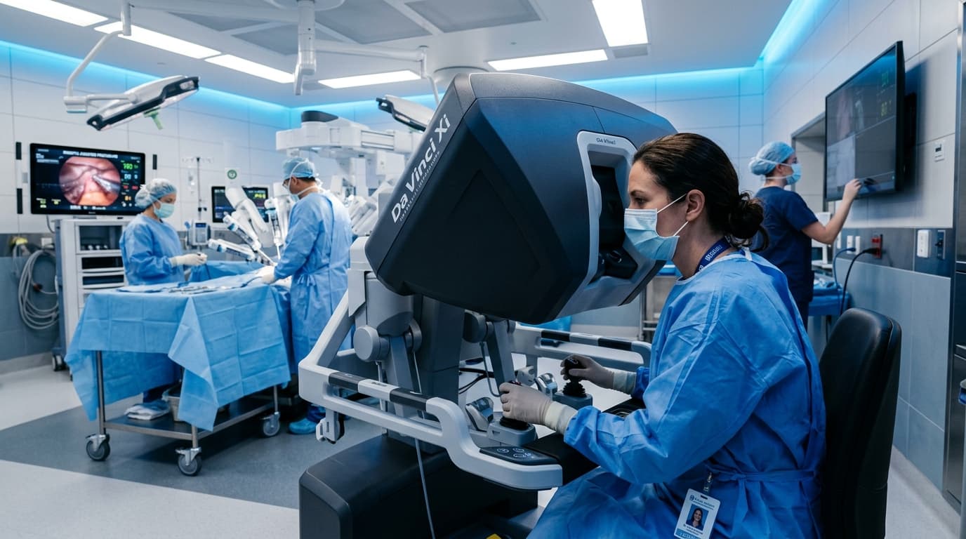
Robotic Surgery in Urology in Rabat: What Patients Should Know Before Deciding
Understand robot-assisted urological surgery, its clinical indications, limits, and essential questions to ask before deciding.

Prostate Cancer: From PSA to MRI, Understanding the Diagnostic Pathway Before Discussing Surgery
An elevated PSA does not automatically mean cancer. Learn how PSA, clinical exam, multiparametric MRI, and targeted biopsy fit together.
Robot-Assisted Radical Prostatectomy: 12 Essential Questions to Ask Before Surgery
Facing prostatectomy? Here are 12 vital questions concerning surgical indication, nerve sparing, continence, erectile function, and follow-up.