
Prostate fusion biopsy: a more accurate diagnosis
Fusion biopsy combines prostate MRI with real-time ultrasound to sample directly from the suspicious lesion. Procedure, approaches and aftercare, explained by Dr Najib Abakka in Rabat.

Dr Najib Abakka
Urological Surgeon in Rabat
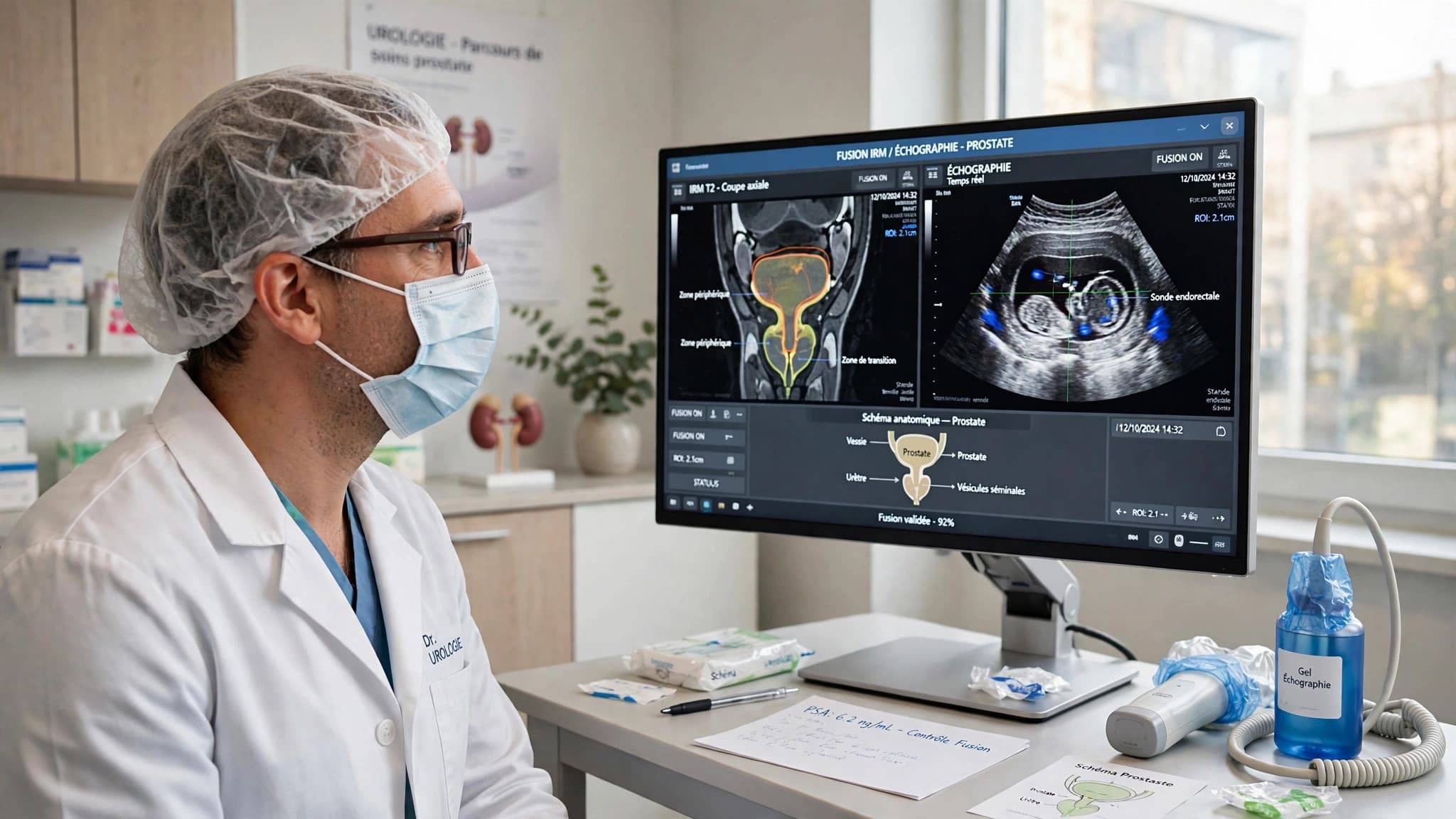
La fusion IRM-échographie permet de prélever directement sur la lésion suspecte.
What is image fusion biopsy?
Image fusion biopsy combines two examinations: a prostate MRI, performed beforehand, and ultrasound, performed in real time during the procedure. Software overlays the two images, so the lesion seen on MRI appears directly on the ultrasound image.
The biopsy needle is then guided precisely onto the target, instead of sampling blindly. The procedure takes place at the clinic, usually under sedation or loco-regional anaesthesia: the patient feels absolutely nothing during the examination.
Why fusion improves diagnosis
MRI identifies suspicious areas of the prostate and grades them on the PI-RADS scale: PI-RADS 1 or 2, low risk; PI-RADS 3, intermediate risk; PI-RADS 4 or 5, high suspicion of significant cancer.
Standard biopsy spreads about twelve samples across the whole prostate without seeing the lesion, so it can miss a cancer. By targeting the lesion, fusion detects significant cancers — those that need treatment — more reliably, and avoids unnecessary biopsies.
When is it recommended?
Fusion biopsy is recommended when PSA is high or rising, when MRI shows a lesion (PI-RADS 3, 4 or 5), during active surveillance, or after a previous negative biopsy while suspicion of cancer remains.
The indication is always decided in consultation, after reviewing the file: PSA, MRI, medical history and current treatments.
How does the procedure go?
After the consultation and the preparation instructions — including which medicines to report, especially blood thinners — the procedure takes place at the clinic, usually under sedation or loco-regional anaesthesia.
The target seen on MRI is displayed on the fused image, then the needle samples it directly several times. Systematic samples are taken as well. The procedure lasts about 30 to 45 minutes, and the patient goes home the same day after a short observation period.
Transperineal or transrectal approach?
Biopsy can be performed through the rectum (transrectal) or through the perineum (transperineal). Dr Abakka favours the transperineal approach: because the needle does not pass through the rectum, the risk of infection after biopsy is very low.
The choice of approach is discussed with each patient according to their anatomy and medical history.
Targeted biopsy or standard biopsy: the difference
Standard biopsy spreads about twelve systematic samples across the whole prostate without visualising the lesion: a localised cancer may simply not be sampled.
With fusion, the needle is guided right onto the target and samples it directly, in addition to systematic samples. Diagnosis is therefore more accurate, especially for significant cancers that need treatment.
After the biopsy
After-effects are usually mild and temporary: a little blood in the urine, stools or semen for a few days, and slight discomfort.
Fever, inability to urinate or heavy bleeding should be reported promptly. Sample analysis results are available within a few days, then explained in consultation together with the appropriate management plan.
Frequently asked questions
Is the procedure painful?
No. Fusion biopsy is performed at the clinic, usually under sedation or loco-regional anaesthesia: the patient feels absolutely nothing during the procedure.
How long does the biopsy take?
About 30 to 45 minutes, then a short observation period before going home the same day.
How should I prepare?
Report all your medicines, especially blood thinners and antiplatelet drugs: the doctor will tell you whether to stop them. Other instructions, including having someone accompany you after sedation, are given before the procedure.
What are the risks?
The most common are mild and temporary: blood in the urine, stools or semen for a few days. Infection is rare, and the transperineal approach favoured by Dr Abakka makes it very unlikely, because the needle does not pass through the rectum.
When are results available?
Sample analysis takes a few days. Results are then explained in consultation, together with the appropriate management plan.
What if the biopsy finds no cancer?
Follow-up continues, with PSA and MRI according to the estimated risk. A persistent suspicious lesion may justify further evaluation.
Related guides & articles
Browse all articles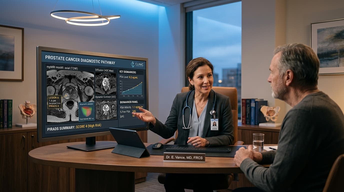
Prostate Cancer: From PSA to MRI, Understanding the Diagnostic Pathway Before Discussing Surgery
An elevated PSA does not automatically mean cancer. Learn how PSA, clinical exam, multiparametric MRI, and targeted biopsy fit together.
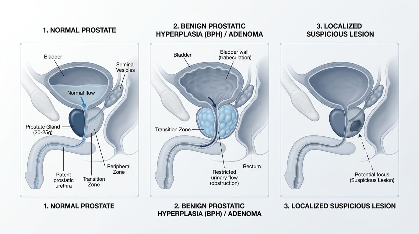
Prostate Adenoma (BPH) or Cancer? Overlapping Symptoms and Distinguishing Tests
Weak stream, nocturnal urination, PSA: benign prostatic hyperplasia and prostate cancer cannot be diagnosed on symptoms alone.

Life After Prostate Surgery: Continence, Sexual Health, PSA, and Follow-Up
What to expect after radical prostatectomy? Catheter care, urinary continence, erectile recovery, pathology results, and PSA monitoring.