
Urinary stone disease: urinary stones
A stone blocking the flow of urine causes renal colic, one of the most intense pains there is. Endoscopic thulium laser treatment shatters it through the natural passages, without any incision.
Dr Najib Abakka
Urological Surgeon in Rabat
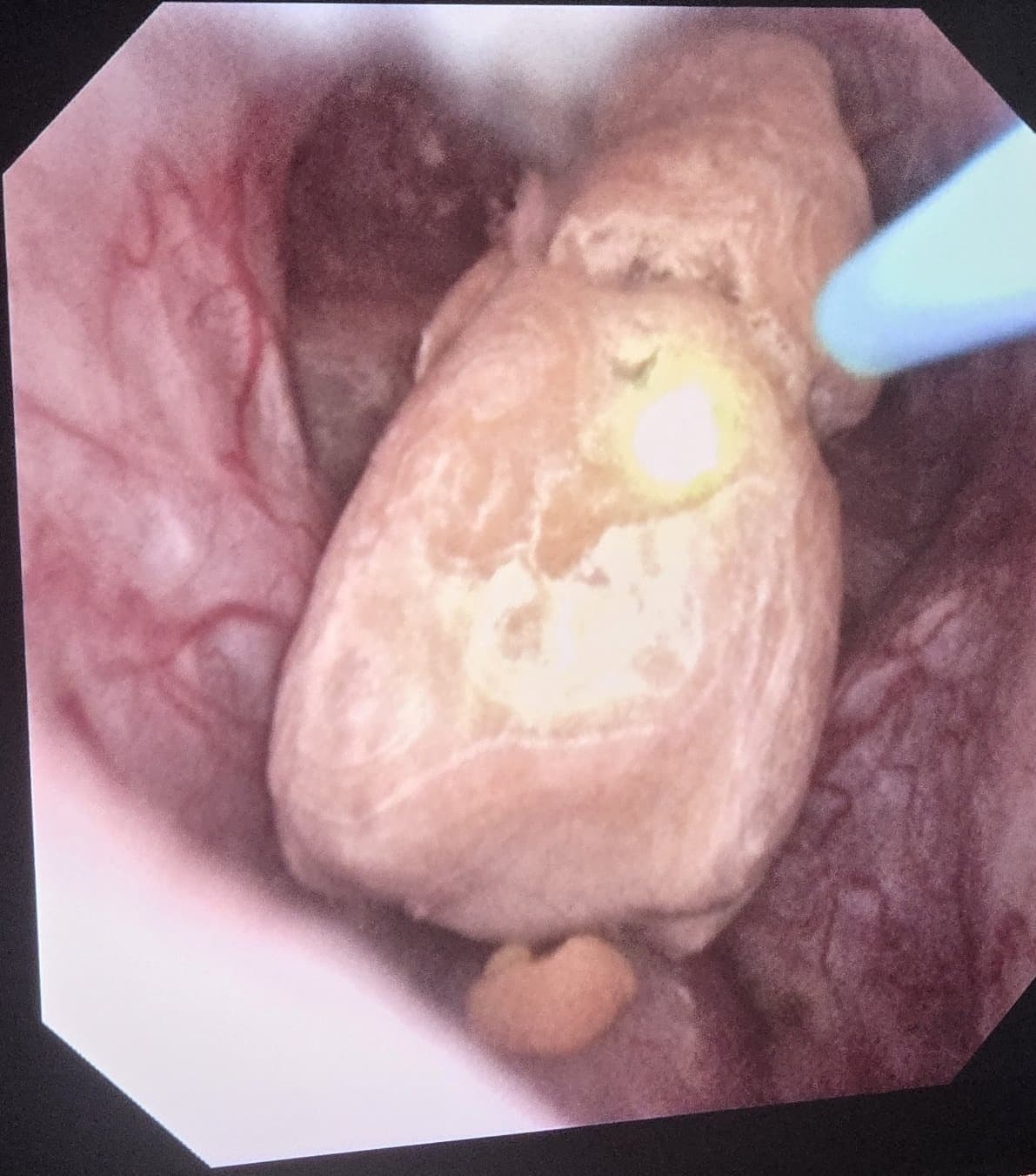
Fragmentation d'un calcul au laser thulium filmée au bloc opératoire (vue endoscopique réelle).
What is a urinary stone?
Urine naturally contains dissolved mineral salts. When they become too concentrated — because you do not drink enough, or for dietary, metabolic or infectious reasons — they crystallise and form a stone. The stone grows in the kidney, then can move into the ureter (the channel connecting the kidney to the bladder), where it gets stuck and triggers pain.
A stone can stay in the kidney without any symptoms, get stuck in the ureter — this is renal colic — or form directly in the bladder. The stone's size, location and hardness determine the treatment.
What are stones made of? Knowing the nature of a stone — thanks to analysis of the recovered fragment — helps tailor prevention. Four main families:
Calcium — The most common: calcium oxalate or phosphate: about 8 stones in 10. Often linked to urine that is too concentrated and to dietary habits.
Uric acid — Common: favoured by acidic urine, gout and excess animal protein. One particular feature: these are the only stones that can sometimes be dissolved with medical treatment.
Infectious — Linked to infections: known as "struvite" stones, they form in the presence of certain urinary infections and can become very large ("staghorn" stones).
Cystine — Rare: of genetic origin (cystinuria), they often appear from childhood or in young adults and tend to recur.
Factors that favour stones:
Drinking too little — this is the leading cause: concentrated urine crystallises more easily, especially in a hot climate.
Diet: excess salt, animal protein and sugar; in some cases, excess foods rich in oxalates (spinach, beetroot, chocolate, very strong tea…).
Recurrent urinary infections.
Background: family history of stones, gout, diabetes, obesity, certain diseases (hyperparathyroidism) or abnormalities of the urinary tract.
Certain medicines and prolonged sedentary behaviour.
Symptoms: from renal colic to the silent stone
Renal colic:
Sudden, intense pain in the back and flank, under the ribs, radiating towards the lower abdomen and the genitals.
Wave-like pain, no position brings relief: the patient is restless, "frantic".
Nausea, sometimes vomiting.
Frequent urge to urinate, burning if the stone is close to the bladder.
Blood in the urine (pink urine), visible or detected on testing.
Other presentations:
A stone that has stayed in the kidney can be completely silent and discovered by chance on an ultrasound scan.
Recurrent urinary infections can be the only sign.
A dull, persistent flank pain, when the stone keeps hampering the flow of urine.
Over time, an obstructed, untreated kidney can silently lose its function — hence the importance of not leaving an obstructing stone untreated.
Pain + fever = absolute emergency. Renal colic accompanied by fever or chills means the urine blocked upstream of the stone is infected: the risk is a serious blood infection (sepsis). Go to the emergency department immediately — the kidney must be drained urgently (double-J stent or nephrostomy), before the stone itself is treated. Same emergency if you cannot urinate at all or if you have only one kidney.
Diagnosis: seeing the stone, measuring it, deciding
CT of the urinary tract (CT urography, most often without contrast injection) is the reference examination: it locates the stone precisely, measures its size and density — an indicator of its hardness — and shows whether the kidney is dilated upstream. These are the elements that make it possible to choose between spontaneous passage, lithotripsy and laser endoscopy.
Kidney and bladder ultrasound: often the first examination, also useful for follow-up (no radiation).
Urine tests: looking for blood and, above all, infection.
Blood tests: kidney function, calcium, uric acid…
Analysis of the stone (or its fragments), essential after a first episode: it identifies its nature and guides prevention of recurrences.
Metabolic work-up (blood and 24-hour urine) in case of recurrent or multiple stones: it looks for the precise cause of stone formation.
Not all stones need surgery. A small ureteral stone (about less than 5 mm) has a good chance of passing on its own with suitable medical treatment — painkillers, plenty of fluids and, if needed, a medicine that helps expulsion. An intervention is considered when the stone is too large, the pain does not settle, there is infection, or the kidney is suffering.
Endoscopic laser treatment
It is now the reference technique for most stones of the kidney, ureter and bladder. With the advent of the thulium laser and of endoscopes that are ever smaller, thinner and more manoeuvrable, flexible ureteroscopy and endoscopic approaches have replaced the older techniques. A thin endoscope — the ureteroscope, rigid or flexible — is introduced through the natural passages (urethra, bladder, then ureter) up to the stone, which the surgeon sees live on screen. A thulium laser fibre a few tenths of a millimetre wide, slipped inside the endoscope, then delivers its energy exactly in contact with the stone.
Two ways of using the laser:
Fragmentation: the laser breaks the stone into fragments, recovered one by one with a tiny basket — one of them is sent for analysis.
Dusting: set differently, the laser reduces the stone to dust, passed spontaneously in the urine in the days that follow.
The advantages:
No incision, no scar: everything passes through the natural passages.
Effective on all types of stones, even the hardest, where external shock waves can fail.
The flexible ureteroscope reaches all the cavities of the kidney, including hard-to-reach areas.
Result checked immediately: at the end of the procedure the surgeon verifies that no significant fragment remains.
Fast recovery and low post-operative pain.
Combined with ever smaller endoscopes, the thulium laser makes it possible to reach and treat stones that were once reserved for much heavier techniques.
The laser used is the thulium laser (thulium fibre laser): it is the one that changed the game. Its energy is absorbed on contact with the stone with millimetric precision that spares the neighbouring tissues; it reduces the stone to particularly fine dust, with very little retropulsion of the stone during firing — which makes dusting more complete, faster and safer, even in the kidney cavities reached by the flexible ureteroscope.
Thulium laser + ever smaller endoscopes: the technique that has replaced ESWL and PCNL. ESWL (extracorporeal shock wave lithotripsy) acts blindly, through the skin: its result is not reliable — failures on hard stones, remaining fragments, sessions to repeat, then painful passage of fragments — with no direct control of what remains. PCNL (percutaneous nephrolithotomy), on the other hand, is very invasive: it requires going through the back and piercing the kidney, with a clearly higher risk of bleeding and complications and a longer hospital stay. By contrast, flexible ureteroscopy and endoscopic approaches pass only through the natural passages, see the stone live and treat it in contact with the thulium laser: a far less invasive technique, with far fewer risks — which is why it has become the reference and has replaced ESWL and PCNL in the great majority of cases.
How does the procedure go?
From the decision CT scan to removal of the stent, the pathway has four steps.
The decision: at the consultation, the CT scan is analysed: size, position and density of the stone. The most suitable technique is chosen with you, and the procedure scheduled (outside emergencies).
Anaesthesia: the procedure is most often performed under general anaesthesia (sometimes spinal anaesthesia), on an empty stomach. You feel nothing during the procedure.
The laser step: the endoscope — ever smaller — travels up through the natural passages to the stone, under visual and X-ray guidance. The thulium laser fibre fragments or dusts it; the fragments are extracted with the basket. Duration: 30 to 60 minutes.
The double-J stent: if necessary, a thin internal tube (a "double-J" stent) is left for a few days between the kidney and the bladder to keep urine flowing safely. It is removed at the consultation, in a few minutes.
The day of the procedure:
Hospital stay: most often day surgery (discharge the same day) or a single night of observation.
Before: a recent urine test is essential — the procedure is postponed if an untreated urinary infection is present. Tell us about your treatments, especially anticoagulants and antiplatelet drugs.
After: you leave with a prescription for painkillers and hydration instructions; a filter may be given to you to collect the fragments and have them analysed.
Resuming activities: light activities and work resume within a few days; intense effort is avoided for one to two weeks, especially if a double-J stent is in place.
Thulium laser, shock waves, surgery: why endoscopy has won out
Flexible thulium laser ureteroscopy, carried by ever smaller endoscopes, has replaced ESWL and PCNL in the great majority of cases: it treats the stone live, through the natural passages, with far fewer risks. ESWL and PCNL keep only residual indications, detailed below.
External lithotripsy (ESWL): shock waves, delivered through the skin, fragment the stone blindly, without seeing it or controlling the result. Indications now very limited: a few moderately sized kidney stones, not very hard and well located. Anaesthesia: sedation or light anaesthesia, sometimes simple premedication. Hospital stay: day surgery, sessions sometimes repeated. Particular features: unreliable result: frequent remaining fragments, sessions often repeated, then sometimes painful passage in the urine; failures on hard stones.
Thulium laser endoscopy: flexible endoscope through the natural passages; the thulium laser fibre fragments or dusts the stone on contact, under direct visual control. First-line technique: ureteral stones, kidney stones (up to about 2 cm, including hard stones or stones after failed ESWL) and bladder stones. Anaesthesia: general anaesthesia most often. Hospital stay: day surgery or one night. Particular features: far less invasive and far less risky: no incision, result checked live, treatment in a single session most often; a temporary double-J stent is sometimes placed.
Percutaneous surgery (PCNL): a tract through the back pierces the kidney to access it directly; the stone is fragmented and aspirated. Remains indicated only for very large kidney stones (over 2 cm) and large staghorn stones. Anaesthesia: general anaesthesia. Hospital stay: generally 2 to 3 nights. Particular features: very invasive technique: the kidney is pierced, with a higher risk of bleeding and complications, and a scar on the back — reserved for very large stone volumes.
Alongside these techniques, some stones pass spontaneously with medical treatment, and uric acid stones can sometimes be dissolved by a treatment that alkalinises the urine. These options are discussed at the consultation according to your CT scan.
After the procedure: recovery and preventing recurrences
Recovery is most often straightforward. Some effects are common and harmless:
Pink urine for a few days.
Mild burning on urination, during the first days.
If a double-J stent is in place: frequent urge to urinate, flank discomfort when passing urine, sometimes a little blood in the urine — all of this disappears when the stent is removed.
Passing dust or micro-fragments can cause small colic-type pains, eased by the prescribed painkillers.
Follow-up includes imaging to check that no fragment remains, analysis of the removed stone and, if needed, a metabolic work-up. It is from these elements that prevention is personalised.
Avoiding recurrence: the rules that matter:
Drink enough to pass more than 2 litres of urine a day — clear urine all day, including in the evening, and more in hot weather or on exertion: this is the most effective measure, for all types of stones.
Reduce salt (it increases calcium elimination in the urine).
Moderate animal protein (meat, offal, processed meats), especially in case of uric acid stones.
Do not cut out dietary calcium (dairy products in normal amounts): contrary to a common belief, a low-calcium diet favours calcium stones.
Depending on the nature of your stone, targeted advice — and sometimes long-term medical treatment — will be added at the consultation.
Seek urgent care if, in the days that follow: fever above 38.5 °C or chills • intense pain not relieved by the prescribed painkillers • inability to urinate • heavy bleeding or clots in the urine • vomiting preventing you from drinking.
Videos: endoscopic treatment in real conditions
Frequently asked questions
Is laser treatment painful?
No during the procedure, performed under general anaesthesia. Afterwards, moderate discomfort is possible: burning on urination during the first days and, if a double-J stent is placed, frequent urge to urinate. Simple painkillers are usually enough.
How long is the double-J stent kept?
A few days to two weeks in most cases. It is removed at the consultation, in a few minutes, using a small flexible endoscope passed through the natural passages — without general anaesthesia. All stent-related discomfort disappears after its removal.
Can all stones be treated with laser?
The very great majority, yes: with the thulium laser and ever smaller flexible ureteroscopes, ureteral stones, kidney stones up to about 2 cm and bladder stones — including the hardest — are treated through the natural passages. It is this technique that has replaced ESWL and, in most cases, PCNL, which is far more invasive. Percutaneous surgery remains useful only for very large kidney stones (over 2 cm, large staghorn stones), sometimes combined with endoscopy. Your CT scan makes it possible to decide before the procedure.
Is the thulium laser dangerous for the kidney or ureter?
No: it is precisely for its safety that the thulium laser has become established. The fibre is very fine and its energy acts only in immediate contact with the stone, under constant visual control, with very little retropulsion of the stone during firing. Through the natural endoscopic passages — without incision or perforation of the kidney — the risks are far lower than with PCNL, and the result is checked live, unlike ESWL. Complications remain rare: urinary infection, or injury to the ureter that may require a double-J stent for longer. The benefit-risk balance is discussed with you at the consultation.
Can the stone come back after treatment?
Yes: treatment removes the stone, but not the cause that made it. Without preventive measures, about one patient in two has a recurrence within 5 to 10 years. Hence the importance of stone analysis, metabolic work-up in case of recurrence, and hygiene measures — starting with drinking enough to pass more than 2 litres of urine a day.
Can I wait for the stone to pass on its own?
For a small ureteral stone without fever or impact on the kidney, monitoring with medical treatment is often offered first. But a stone that does not progress after a few weeks, that makes the kidney suffer or that becomes infected must be treated: an obstructed kidney can lose its function without making a sound. Imaging follow-up makes it possible not to exceed a reasonable time limit.
Related guides & articles
Browse all articles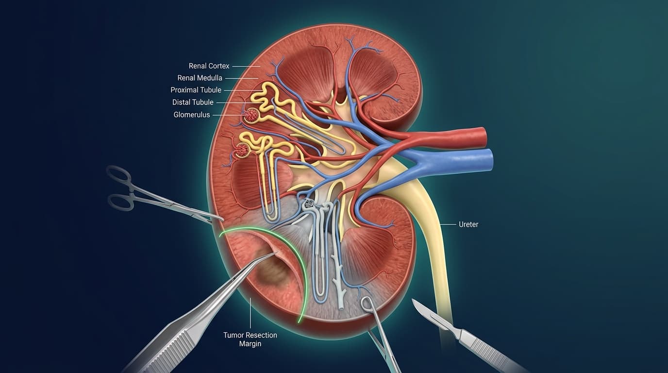
Kidney Cancer: When Can Partial Nephrectomy Preserve the Healthy Kidney?
When can a renal mass be removed while saving the remainder of the kidney? Clinical indications, renal function preservation, and minimally invasive options.
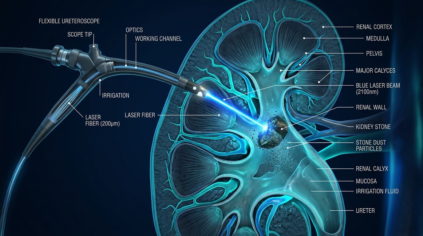
Kidney Stones: When is Laser Ureteroscopy Recommended?
How does laser ureteroscopy work, for which stones is it recommended, and what should you know about JJ stents and recovery?
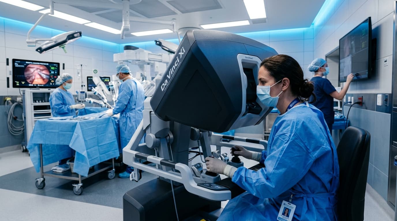
Robotic Surgery in Urology in Rabat: What Patients Should Know Before Deciding
Understand robot-assisted urological surgery, its clinical indications, limits, and essential questions to ask before deciding.